My Favorite Tissue: The Human Ear!
The ear is one of the mammalian sensory organs. Our senses help us to interact with the world around us. The ear allows us to hear sounds (from a lecture to our favorite song) and also plays an important role in our balance!
Figure 1: The Ear.
http://audaud.com/2005/03/the-system-is-the-hero/
Anatomy of the Human Ear
The ear is made up of three main parts:
1) External ear: The external ear is the part of the ear which is visible. It is known as the pinna or ear lobe and is responsible for collecting sound.
** Separating the external ear from the middle ear is the tympanic membrane (aka ear drum). An ear canal leads the pinna to the ear drum. **
2) Middle Ear: The middle ear contains 3 small bones( incus, mallus and stapes) collectively known as the ossicles. These small bones move the sound from the ear drum to the inner ear.
3) Inner Ear: Contains the round window and the oval window. Also contains two organs, the semicircular canals and the cochlea. The cochlea is responsible for hearing. It converts hair movements into electrical impulses which go to the brain via the auditory nerve. The semicircular canals help us in maintaining our balance.

Figure 2: Anatomy of the Human Ear
Function of the Ear:
There are two main functions of the ear:
A) Enable us to hear
B) Balance
A) How do we hear a song on the radio, a baby cry or a bird sing??
There is a stepwise set of events that occur in order for us to hear:
1. The pinna collects sound waves and they enter the ear canal in order to reach the ear drum.
2. The ear drum vibrates as well as the ossicles.
3. These vibrations also causes the fluid in the inner ear to vibrate.
4. Vibration of the fluid causes hair cells to move and the movement is turned into electrical signals.
5. The signals go through the auditory nerve and eventually to the brain where the signal gets interpreted.
6. Hearing results!
B) We often don't think of our ears as keeping us in balance. However, the vestibular system in our inner ear consisting of semicircular canals, saccule, and utricle sends information to the brain about balance and if an abnormal signal is received the body tries to adjust by changing posture or eye movements for example.

Figure 3: Balance
http://www.personal-nutrition-guide.com/ideal-weight.html

Figure 4: Semicircular Canal
Histology: (Young et al., 2006).
External Ear: The pinna is made of elastic and fibrocartilage covered in skin. Cartilage is made up of cells called chondrocytes and an extracellular matrix (fibers and ground substance). The fibers are elastic fibers which make the pinna flexible. The pinna is attached to the head by muscles and ligaments.The ear canal from the pinna to the tympanic membrane (Meatus acusticus extera) has a lateral component that is a cartilage tube, lined with squamous epithelium. It also has hair follicles, and subaceous glands. Modified sweat glands called ceruminous glands produce ear wax! The medial
portion of the canal is bony and is lined with stratified squamous epithelium (Baguley et al., 2013).
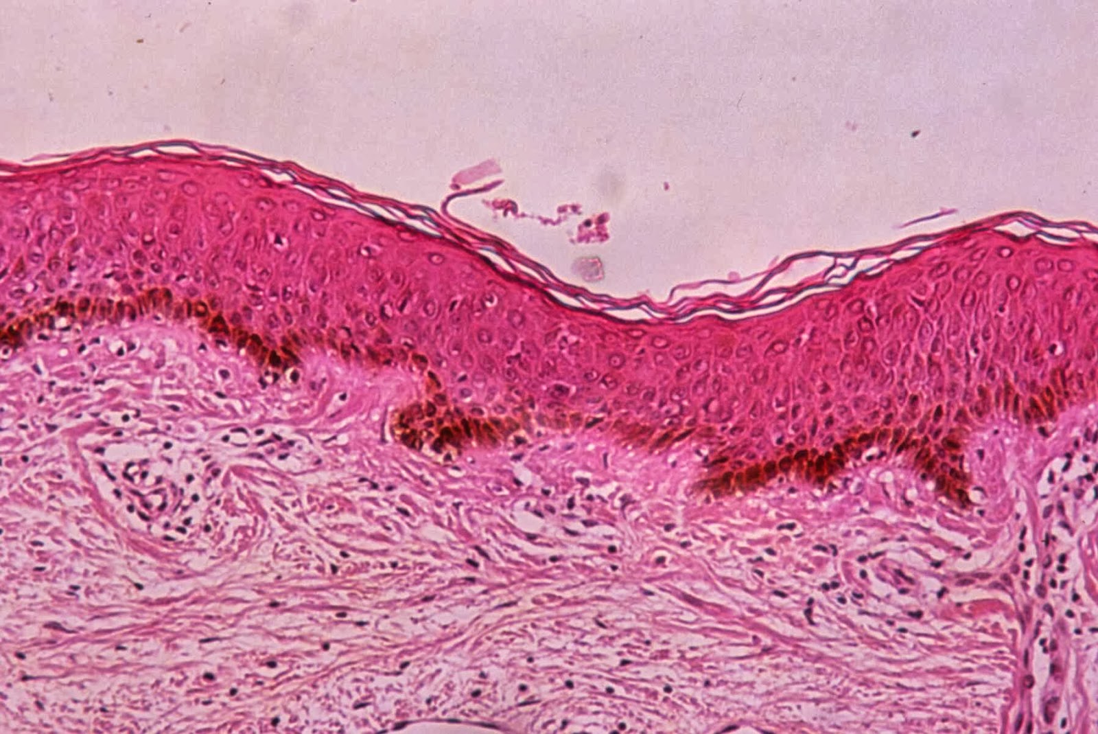
Figure 5: Pinna
http://pinna.hawkelibrary.com/Normal-Anatomy/1_4?full=1
Tympanic Membrane( Ear Drum): The ear drum is made up of three layers. The external cuticular layer is continuous with the epithelium of the ear canal and is covered in hairless skin. The thin dermis has fibroblasts and a vascular network. The intermediate fibrous layer is a layer of fibers that are made up of type II and III collagen and a small amount of coarse collagen type I fibers. The inner part of the ear drum is known as the inner mucous layer. It is a single layer of cuboidal cells. It has a thin lamina propria with a blood supply separate from the external cuticular layer.

Figure 6: Ossicles
http://www.kids-ent.com/newkids800/ossicles.html
Inner Ear: The inner ear is composed of the bony labyrinth (semicircular canals, vestible and cochlea). and the membraneous labyrinth which runs inside the bony labyrinth and is fluid filled. The inner ear also contains the cochlea. The cochlea is a tube shaped structure that is shaped like a snail shell. It has two outer canals (the vestibular canal and the tympanic canal). A central canal called the cochlear duct runs between the two outer canals. Sensory hair cells are located in the columnar epithelium on the floor of the cochlear duct. The hair cells and supporting cells make up the Organ of Corti which is responsible for hearing.

Figure 7: Organ of Corti
http://bio1903.nicerweb.com/Locked/media/ch50/cochlea.html
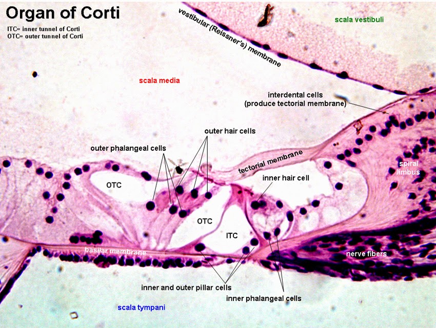
Figure 8: Organ of Corti
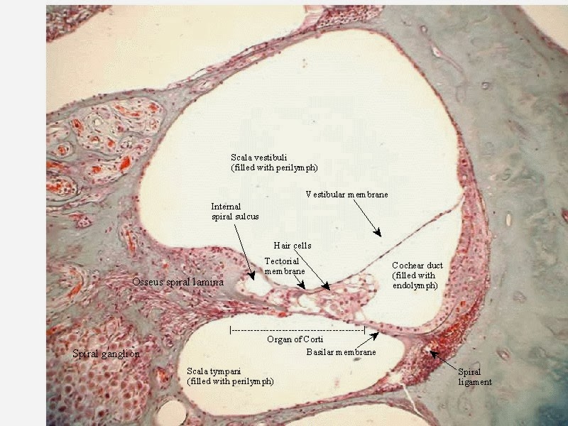
Figure 9: Cochlear Duct
Pathology:
There are many different diseases that can affect the ear and also different types of deafness.
Conductive Deafness: Occurs when something interferes with sound conduction from the outer to the middle ear, therefore the sound is affected before it even reaches the cochlea. It can be caused by many different things such as wax in the external ear, inflammation of the middle ear or from an inherited disease called otosclerosis(when the ossicles fuse together) (Young et al., 2006).
Sensory Neural Deafness: (Young et al., 2006) This occurs when the sensory receptors in the inner ear or the auditory nerve is damaged. There are different cases that cause this type of deafness:
1) Children can be born with it (due to low O2 during birth or an intrauterine infection).
2) Noise exposure
3) Age- as you get older you may lose high frequency hearing.
Congenital Hearing Loss: An example of this type of hearing loss is when Cytomegalovirus(CMV) is transmitted from mother to fetus. CMV is a herpes virus that can occur when you come into contact with blood, urine, and breast milk. Sensorineural hearing loss is something that commonly happens when infected with this disease.(Beyea et al., 2012).
Sudden Sensorineural Hearing Loss: This type of hearing loss could be due to viral invasion of the fluid and tissues of the cochlea or viral invasion of the cochlear nerve. It is categorized as losing at least 30 dB of hearing in 3 or more consecutive frequencies in less than 3 days (Beyea et al., 2012).
Meniere's Disease: (Yamane et al., 2010). This disease occurs when there is an abnormality in the inner ear. Usually due to a disturbance of fluid (disturbance of the flow of endolymph).This disease has many symptoms including:
- loss of balance
-dizziness
-tinnitus (ringing in the ear).
-some hearing loss
Acute Otitis Media: This is a disease caused by inflammation of the lining of the middle ear and ear drum which can cause swelling. It is similar to an infection of the nose and sinuses. If the swelling becomes too great the ear drum may burst which can relieve pain. Sometimes this disease recovers without treatment and sometimes antibiotics are used (Lambert & Soham, 2013).
Treatment:
There are many different treatments which can be used for the different diseases/ deafness of the ear.
Hearing aids are a common treatment for hearing loss. They are a small electronic device which can fit in your ear or behind it. Hearing aids make sounds louder so they are used when hearing loss is mild to severe. If a hearing aid is not enough another treatment such as cochlear implants may be used. (http://www.hearinglosseducation.com/treatments/hearing-aids).
One of the common treatments for recurring acute otitis media (AOM) is Myringotomy ear tubes. They are placed through the ear drum to help fluid behind the ear drum drain so the ear can work properly. They are placed in the ear if there is more than 3 cases of AOM in 6 months or 4 cases in a year. This is very common in children (Lambert & Soham, 2013).
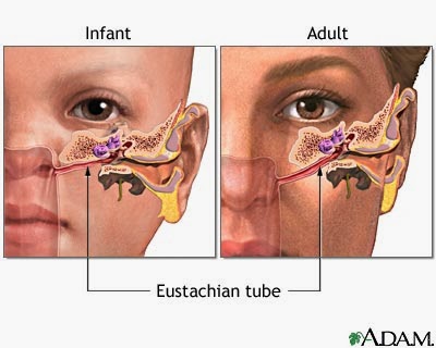
Figure 10: Eustachian Tubes
http://www.healthcentral.com/cold-flu/h/sinus-infection-tubes-in-ear-pictures.html
Another common treatment is the use of cochlear implants. They are an electronic device that are surgically implanted. Used in patients who are deaf due to damage to hair cells. They are often used in children with sensorineural hearing loss. Should be implanted at a young age (preferably before age 2) to increase the chances of reaching normal milestones ( Lee-Suk et al., 2010).
Interesting Facts: Did you Know???
http://www.calmear.com/index.php?option=com_content&view=article&id=6&Itemid=8
 Figure 11: Interesting Facts
http://www.calmear.com/index.php?option=com_content&view=article&id=6&Itemid=8
* Ear piercing is a common cosmetic and cultural practice.

Figure 12: Ear Piercing
http://www.sarahhurst.co.uk/ear-piercing-brighton/
* Your ear never stops working! Even when you sleep your ear still hears sounds, but the brain blocks it out.
* The skin of the ear canal grows 1.3 inches per year and then falls off. You get a new ear canal each year!! If it didn't fall off, by the time you were 20 you would have 20 feet of ear canal hanging out of your ear!!
*Wearing headphones creates a good environment for bugs. Bacteria increases by 700X in each hour!!
* Many people who are deaf or hard of hearing use sign language to communicate. American Sign Language uses body language, facial expressions and signs by the hands to communicate.(http://www.nidcd.nih.gov/health/hearing/pages/asl.aspx#2)
*Ossicles (incus, malleus, and stapes) are the three smallest bones in the human body. The three of them can fit on a penny!

Figure 13: Ossicles on a Penny
http://www.sharinginhealth.ca/biology/ear.html
References:
Anonymous. 2010. Calm Ear. Available at:
http://www.calmear.com/index.php?option=com_content&view=article&id=6&Itemid=8
Baguley, D., Anderson G., Mcferran, D., & McKenna L. 2013. Tinnitus: A Multidisciplinary Approach. 2nd Edition.
Beyea, J.A., Agrawal, S.K., & Parnes, L.S. 2012. Recent Advances in Viral Inner Ear Disorders. Current Opinion in Otolarynology Head and Neck Surgery 20: 404-408.
Hearing Loss Education Center. 2013. Types of Hearing Loss. Available at:
http://www.hearinglosseducation.com/types-of-hearing-loss
Lambert, E. & Soham, R. 2013. Otitis Media and Ear Tubes. Pediatric Clinics of North America 60.
Lee-Suk, K., Sung-Wook, J., Young-Mee, L. & Jeong-Seo, K. 2010. Cochlear Implantation in Children. Auris Nasus Larrynx 37:6-17.
National Institute on Deafness and Other Communication Disorders. 2012. American Sign Language. Available at: http://www.nidcd.nih.gov/health/hearing/pages/asl.aspx#2
University of California, Irvine. School of Medicine. Department of Otolaryngology. Anatomy of the Ear. Available at:
http://www.ent.uci.edu/clinical-specialties/facial-nerve-and-skull-base-surgery/ear-anatomy
Yamane, H., Takayama, M., Sunami, K., Sakamoto, H., Imoto, T., & Anniko, M. 2010. Blockage of Reuniting Duct in Meniere's Disease. Acta Oto-Laryngologica 130: 233-239.
Young, B., Low, J.S., Stevens, A., & Heath, J.W. 2006. Wheater's Functional Histology. Atext and Color Atlas 5th Edition.